Cell Image Analysis Technology Applications
Hematopoietic Stem Cell Tracking
| We present a method for robustly detecting hematopoietic stem cells (HSCs) in phase contrast microscopy images. HSCs appear to be easy to detect since they typically appear as round objects. However, when HSCs are touching and overlapping, showing the variations in shape and appearance, standard pattern detection methods, such as Hough transform and correlation, do not perform well. The proposed method exploits the output pattern of a ring filter bank applied to the input image, which consists of a series of matched filters with multiple-radius ring-shaped templates. By modeling the profile of each filter response as a quadratic surface, we explore the variations of peak curvatures and peak values of the filter responses when the ring radius varies. The method is validated on thousands of phase contrast microscopy images with different acquisition settings, achieving 96.5% precision and 94.4% recall. | ||||
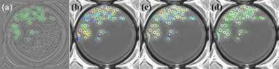
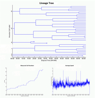
(a)Filtering: Output of ring filter with radius of 12 pixels and the corresponding candidates. (b)Cell Candidates: all candidates from ring filters with radii between 9 and 15 pixels. (c)Cell detection result after false positie elimination. (d)Cell tracking by association. (e)Linage trees and statistics(number of cells and cells speed). |
 |
|||
|
Two fundamental barriers hinder the development of improved HSC therapy: the lack of cost effective strategies to boost the number of HSCs in vitro; and the lack of understanding of the conditions for controlled differentiation of HSCs into various types of tissues. To overcome these barriers, novel computational toolsets are required to reliably quantify the behaviors of HSCs in population environments, as they divide into daughter cells. We have developed a robust method for the tracking of HSCs in phase contrast images for implementing such toolsets. | ||||
|
Detecting Hematopoietic Stem Cells (HSC) imaged with phase contrast microscopy |
Tracking Hematopoietic Stem Cells (HSC) imaged with phase contrast microscopy |